The number of chromosomes in different types of organisms. What is a chromosome? Set of chromosomes
Poor ecology, life in constant stress, priority of career over family - all this has a bad effect on a person’s ability to bear healthy offspring. Sadly, about 1% of babies born with serious chromosome abnormalities grow up mentally or physically retarded. In 30% of newborns, deviations in the karyotype lead to the formation of congenital defects. Our article is devoted to the main issues of this topic.
The main carrier of hereditary information
As is known, a chromosome is a certain nucleoprotein (consisting of a stable complex of proteins and nucleic acids) structure inside the nucleus of a eukaryotic cell (that is, those living beings whose cells have a nucleus). Its main function is the storage, transmission and implementation of genetic information. It is visible under a microscope only during processes such as meiosis (division of a double (diploid) set of chromosome genes during the creation of germ cells) and mycosis (cell division during the development of the organism).
As already mentioned, a chromosome consists of deoxyribonucleic acid (DNA) and proteins (about 63% of its mass) on which its thread is wound. Numerous studies in the field of cytogenetics (the science of chromosomes) have proven that DNA is the main carrier of heredity. It contains information that is subsequently implemented in a new organism. This is a complex of genes responsible for hair and eye color, height, number of fingers, etc. Which genes will be passed on to the child are determined at the time of conception.
Formation of the chromosome set of a healthy organism
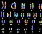
A normal person has 23 pairs of chromosomes, each of which is responsible for a specific gene. There are 46 in total (23x2) - how many chromosomes a healthy person has. We get one chromosome from our father, the other is passed on from our mother. The exception is 23 pairs. It is responsible for the gender of a person: female is designated as XX, and male as XY. When the chromosomes are in a pair, this is a diploid set. In germ cells they are separated (haploid set) before being subsequently united during fertilization.
The set of characteristics of chromosomes (both quantitative and qualitative) examined within one cell is called a karyotype by scientists. Violations in it, depending on the nature and severity, lead to the occurrence of various diseases.
Deviations in the karyotype
When classified, all karyotype abnormalities are traditionally divided into two classes: genomic and chromosomal.
With genomic mutations, an increase in the number of the entire set of chromosomes, or the number of chromosomes in one of the pairs, is noted. The first case is called polyploidy, the second - aneuploidy.
Chromosomal abnormalities are rearrangements both within and between chromosomes. Without going into scientific jungle, they can be described as follows: some sections of chromosomes may not be present or may be doubled to the detriment of others; The sequence of genes may be disrupted, or their location may be changed. Disturbances in structure can occur in every human chromosome. Currently, the changes in each of them are described in detail.
Let us take a closer look at the most well-known and widespread genomic diseases.
Down syndrome
It was described back in 1866. For every 700 newborns, as a rule, there is one baby with a similar disease. The essence of the deviation is that a third chromosome is added to the 21st pair. This happens when the reproductive cell of one of the parents has 24 chromosomes (with double 21). A sick child ends up with 47 chromosomes - that's how many chromosomes a Down person has. This pathology is facilitated by viral infections or ionizing radiation suffered by parents, as well as diabetes.
Children with Down syndrome are mentally retarded. Manifestations of the disease are visible even in appearance: an overly large tongue, large, irregularly shaped ears, a skin fold on the eyelid and a wide bridge of the nose, whitish spots in the eyes. Such people live on average forty years, because, among other things, they are susceptible to heart disease, problems with the intestines and stomach, and undeveloped genitals (although women may be capable of childbearing).
The older the parents are, the higher the risk of having a sick child. Currently, there are technologies that make it possible to recognize a chromosomal disorder at an early stage of pregnancy. Older couples need to undergo a similar test. It will not hurt young parents if one of them has had Down syndrome in their family. The mosaic form of the disease (the karyotype of some cells is damaged) is formed already at the embryonic stage and does not depend on the age of the parents.
Patau syndrome
This disorder is trisomy of the thirteenth chromosome. It occurs much less frequently than the previous syndrome we described (1 in 6000). It occurs when an extra chromosome is attached, as well as when the structure of chromosomes is disrupted and their parts are redistributed.
Patau syndrome is diagnosed by three symptoms: microphthalmos (reduced eye size), polydactyly (more fingers), cleft lip and palate.
The infant mortality rate for this disease is about 70%. Most of them do not live to be 3 years old. In individuals susceptible to this syndrome, heart and/or brain defects and problems with other internal organs (kidneys, spleen, etc.) are most often observed.
Edwards syndrome
Most babies with 3 eighteenth chromosomes die soon after birth. They have pronounced malnutrition (digestive problems that prevent the child from gaining weight). The eyes are set wide and the ears are low. Heart defects are often observed.
conclusions
To prevent the birth of a sick child, it is advisable to undergo special examinations. The test is mandatory for women giving birth after 35 years of age; parents whose relatives were exposed to similar diseases; patients with thyroid problems; women who have had miscarriages.
Containing genes. The name "chromosome" comes from the Greek words (chrōma - color, color and sōma - body), and is due to the fact that when cells divide, they become intensely colored in the presence of basic dyes (for example, aniline).
Many scientists, since the beginning of the 20th century, have thought about the question: “How many chromosomes does a person have?” So, until 1955, all the “minds of humanity” were convinced that the number of chromosomes in humans is 48, i.e. 24 pairs. The reason was that Theophilus Painter (Texas scientist) incorrectly counted them in preparative sections of human testes, according to a court decision (1921). Subsequently, other scientists, using different calculation methods, also came to this opinion. Even after developing a method for separating chromosomes, the researchers did not challenge Painter’s result. The error was discovered by scientists Albert Levan and Jo-Hin Thio in 1955, who accurately calculated how many pairs of chromosomes a person has, namely 23 (more modern technology was used to count them).
Somatic and germ cells contain a different chromosome set in biological species, which cannot be said about the morphological characteristics of chromosomes, which are constant. have a doubled (diploid set), which is divided into pairs of identical (homologous) chromosomes, which are similar in morphology (structure) and size. One part is always of paternal origin, the other of maternal origin. Human sex cells (gametes) are represented by a haploid (single) set of chromosomes. When an egg is fertilized, haploid sets of female and male gametes are united in one zygote nucleus. In this case, the double dialing is restored. It is possible to say with accuracy how many chromosomes a person has - there are 46 of them, with 22 pairs of them being autosomes and one pair being sex chromosomes (gonosomes). Sexes have differences - both morphological and structural (gene composition). In a female organism, a pair of gonosomes contains two X chromosomes (XX-pair), and in a male organism, one X- and Y-chromosome (XY-pair).
Morphologically, chromosomes change during cell division, when they double (with the exception of germ cells, in which duplication does not occur). This is repeated many times, but no change in the chromosome set is observed. Chromosomes are most noticeable at one of the stages of cell division (metaphase). During this phase, the chromosomes are represented by two longitudinally split formations (sister chromatids), which narrow and unite in the area of the so-called primary constriction, or centromere (an obligatory element of the chromosome). Telomeres are the ends of a chromosome. Structurally, human chromosomes are represented by DNA (deoxyribonucleic acid), which encodes the genes that make up them. Genes, in turn, carry information about a specific trait.
Individual development will depend on how many chromosomes a person has. There are such concepts as: aneuploidy (change in the number of individual chromosomes) and polyploidy (the number of haploid sets is greater than the diploid one). The latter can be of several types: loss of a homologous chromosome (monosomy), or appearance (trisomy - one extra, tetrasomy - two extra, etc.). All this is a consequence of genomic and chromosomal mutations, which can lead to pathological conditions such as Klinefelter syndrome, Shereshevsky-Turner syndrome and other diseases.
Thus, only the twentieth century gave answers to all questions, and now every educated inhabitant of planet Earth knows how many chromosomes a person has. The sex of the unborn child depends on the composition of the 23 pairs of chromosomes (XX or XY), and this is determined during fertilization and the fusion of the female and male reproductive cells.
Chromosome is a thread-like structure containing DNA in the cell nucleus, which carries genes, units of heredity, arranged in a linear order. Humans have 22 pairs of regular chromosomes and one pair of sex chromosomes. In addition to genes, chromosomes also contain regulatory elements and nucleotide sequences. They house DNA-binding proteins that control DNA functions. Interestingly, the word "chromosome" comes from the Greek word "chrome", meaning "color". Chromosomes received this name due to the fact that they have the peculiarity of being colored in different tones. The structure and nature of chromosomes vary from organism to organism. Human chromosomes have always been a subject of constant interest to researchers working in the field of genetics. The wide range of factors that are determined by human chromosomes, the abnormalities for which they are responsible, and their complex nature have always attracted the attention of many scientists.
Interesting facts about human chromosomes
Human cells contain 23 pairs of nuclear chromosomes. Chromosomes are made up of DNA molecules that contain genes. The chromosomal DNA molecule contains three nucleotide sequences required for replication. When chromosomes are stained, the banded structure of mitotic chromosomes becomes apparent. Each strip contains numerous DNA nucleotide pairs.
Humans are a sexually reproducing species with diploid somatic cells containing two sets of chromosomes. One set is inherited from the mother, while the other is inherited from the father. Reproductive cells, unlike body cells, have one set of chromosomes. Crossing over between chromosomes leads to the creation of new chromosomes. New chromosomes are not inherited from either parent. This accounts for the fact that not all of us exhibit traits that we receive directly from one of our parents.
Autosomal chromosomes are assigned numbers from 1 to 22 in descending order as their size decreases. Each person has two sets of 22 chromosomes, an X chromosome from the mother and an X or Y chromosome from the father.
An abnormality in the contents of a cell's chromosomes can cause certain genetic disorders in people. Chromosomal abnormalities in people are often responsible for the occurrence of genetic diseases in their children. Those who have chromosomal abnormalities are often only carriers of the disease, while their children develop the disease.

Chromosomal aberrations (structural changes in chromosomes) are caused by various factors, namely deletion or duplication of part of a chromosome, inversion, which is a change in the direction of a chromosome to the opposite, or translocation, in which part of a chromosome is torn off and attached to another chromosome.
An extra copy of chromosome 21 is responsible for a very well known genetic disorder called Down syndrome.
Trisomy 18 results in Edwards syndrome, which can cause death in infancy.
Deletion of part of the fifth chromosome results in a genetic disorder known as Cri-Cat Syndrome. People affected by this disease often have mental retardation and their crying in childhood resembles that of a cat.
Disorders caused by sex chromosome abnormalities include Turner syndrome, in which female sexual characteristics are present but characterized by underdevelopment, as well as XXX syndrome in girls and XXY syndrome in boys, which cause dyslexia in affected individuals.
Chromosomes were first discovered in plant cells. Van Beneden's monograph on fertilized roundworm eggs led to further research. August Weissman later showed that the germ line was distinct from the soma and discovered that cell nuclei contained hereditary material. He also suggested that fertilization leads to the formation of a new combination of chromosomes.
These discoveries became cornerstones in the field of genetics. Researchers have already accumulated a significant amount of knowledge about human chromosomes and genes, but much remains to be discovered.
Video
Cell nucleus
Core(lat. nucleus, Greek karyon) is the most important component of a eukaryotic cell.
The kernel performs two main functions:
- storage and reproduction of genetic information;
- regulation of metabolic processes occurring in the cell, ensuring its normal functioning.
The nucleus contains more than 90% of the DNA of the entire cell.
Most cells have one nucleus. Some cells may contain 2 nuclei (in ciliates these are a macronucleus and a micronucleus).
 In eukaryotic organisms, there are cells that do not have nuclei, but their life span is short (mature red blood cells live on average 125 days). Multinucleated cells (striated muscle fibers, fungal cells) are also known.
Multinucleated cells of striated muscle tissue
The nucleus is most often located in the center of the cell, and only in plant cells with a central vacuole - in the parietal protoplasm.
It can be of various shapes: round, ovoid, horseshoe-shaped, segmented (rarely), elongated, spindle-shaped, etc.


Round nucleus Horseshoe-shaped (bean-shaped) nucleus
The core consists of:
- nucleoplasm;
- chromatin (chromosomes);
- nucleoli;
- the nuclear membrane, which passes into part of the endoplasmic reticulum.

Nuclear envelope
The core is surrounded by a shell consisting of two membranes with a structure typical of all membranes.
 The outer nuclear membrane is covered with ribosomes and passes directly into the channels of the endoplasmic reticulum (endoplasmic reticulum). The inner membrane is smooth and is in contact with the chromosomal material of the nucleus. The membranes are separated from each other perinuclear space
.
The outer nuclear membrane is covered with ribosomes and passes directly into the channels of the endoplasmic reticulum (endoplasmic reticulum). The inner membrane is smooth and is in contact with the chromosomal material of the nucleus. The membranes are separated from each other perinuclear space
.
The thickness of such a double-membrane nuclear envelope is 30 nm. It is permeated with many pores that provide transport of mRNA, tRNA, ATP, enzymes, ions and other substances.
Despite the active exchange between the nucleus and the cytoplasm, the nuclear envelope creates the possibility of the existence of a special internal environment in the nucleus.
Nuclear pores
 The nuclear envelope is penetrated by numerous openings - pores, formed by the fusion of two nuclear membranes. These holes are filled with globular and fibrillar structures. The set of nuclear pores and these structures is called nuclear pore complex
.
The nuclear envelope is penetrated by numerous openings - pores, formed by the fusion of two nuclear membranes. These holes are filled with globular and fibrillar structures. The set of nuclear pores and these structures is called nuclear pore complex
.
Through the pores, substances are exchanged between the nucleus and the cytoplasm. RNA and ribosomal subunits leave the nucleus into the cytoplasm, and the nucleotides necessary for the assembly of RNA, enzymes and other substances that ensure the activity of nuclear structures enter the nucleus.
The number of nuclear pores depends on the metabolic activity of cells: the higher the synthetic processes in cells, the more pores per unit surface of the cell nucleus.
Nuclear juice
 Karyoplasm
, or nucleoplasm
- the fluid contained in the cell nucleus in which all processes occur.
Karyoplasm
, or nucleoplasm
- the fluid contained in the cell nucleus in which all processes occur.
Nuclear juice consists of:
- liquid part;
- nuclear matrix (a kind of framework that penetrates the nuclear juice - strands consisting of acidic proteins);
- various inclusions.
The liquid part is similar in composition to the corresponding component of the cytoplasm: it also contains enzymes, ribosomal and structural proteins of chromosomes, free nucleotides, amino acids and other intermediate products of cell metabolism.
Nucleolus
 A dense round body, consisting of rRNA and ribosomes at different stages of formation, immersed in nuclear juice. In the nuclei of different cells and in the nucleus of the same cell, depending on its functional state, the number of nucleoli can vary from 1 to 5 - 7 or more.
Nucleoli are present only in non-dividing nuclei. During mitosis, they disappear, and then reappear around the region of the chromosome (gene) in which the rRNA structure is encoded. This gene is called the nucleolar organizer (NO). It is where rRNA synthesis occurs.
In addition to rRNA synthesis, ribosomal subunits are synthesized in the nucleolus.
Chromatin(Greek chroma- color, staining) are called DNA-protein complexes.
DNA, located in the cell nucleus, contains information about all the characteristics of an organism. The total DNA length of 46 human chromosomes is 2 meters. However, all of it is packaged in the cell nucleus thanks to special proteins - histones .
Histones- proteins in the nuclei of eukaryotic cells that are part of complexes with DNA. Histones are involved in maintaining and changing the structure of chromosomes at different stages of the cell cycle, as well as in the regulation of gene activity.
There are five types of histones: H1 (very rich in lysine), H2a and H2b (rich in lysine), H3 (rich in arginine) and H4 (rich in glycine and arginine).
The basic unit of chromatin packaging is the nucleosome. It consists of a DNA double helix that wraps 1.75 times around a complex of eight histones.
In chromatin, DNA is presented as a continuous double-stranded thread from one nucleosome to another. Nucleosomes are separated by equal sections of DNA that does not contact the histone complexes. This structure in micrographs looks like beads on a string.
Between divisions, the DNA molecules in the cell are in a despiralized state; it is almost impossible to see them with a light microscope. In a cell preparing to divide, DNA molecules double, spiral, shorten and acquire a compact shape, which makes them visible. In this state, the complex of DNA and proteins is called chromosomes . A chromosome is a continuous strand of chromatin arranged in a specific way.
Chromosomes
Thread-like bodies consisting of DNA molecules contained in the nucleus of a eukaryotic cell - chromosomes (Greek. chroma- color, soma- body). Chromosomes can consist of one or more identical DNA molecules (2, 4, 8, etc.) connected in the region of the primary constriction. The terminal sections of chromosomes (telomeres) protect their ends from sticking together.

The sizes of chromosomes in different organisms differ from each other. Thus, the length of chromosomes can vary from 0.2 to 50 microns. The smallest chromosomes are found in some protozoa and fungi. The longest ones are found in some orthopteran insects, in amphibians and in Liliaceae. The length of human chromosomes ranges from 1.5 to 10 microns.
 Centromere(lat. сentrum, Greek kentron- center and meros- part, lobe) - an area in the area of the primary constriction to which the filaments of the spindle are attached during cell division.The centromere divides the chromosome into two arms of equal or different lengths.
A change in the position of the centromere in a particular chromosome serves as a criterion for identifying chromosomal rearrangements.
Depending on the location of the centromere, four types of chromosome structure are distinguished: 
- metacentric (having arms of equal length);
- submetacentric (with arms of unequal length);
- acrocentric (with a very short second arm);
- telocentric (one shoulder is missing).
In all somatic cells of any plant or animal organism, the number of chromosomes is the same.
Sex cells always contain half as many chromosomes as somatic cells of a given type of organism.
Number of chromosomes in different types of living organisms
| Species name | Number of chromosomes in somatic cells |
|---|---|
| Human | 46 |
| Ash | 46 |
| Gorilla | 48 |
| Buffalo | 48 |
| Chimpanzee | 48 |
| Potato | 48 |
| Black pepper | 48 |
| Dog | 78 |
| Chicken | 78 |
| Cat | 38 |
| Rape | 38 |
| Fox | 38 |
| Guinea pig | 64 |
| Horse | 64 |
| Dove | 16 |
| Onion | 16 |
| Red Ribes | 16 |
| Honey bee | 32 |
| Cherry | 32 |
| House mouse | 40 |
| Cow | 60 |
| Potato | 44 |
| Crayfish | 116 |
| Drosophila fruit fly | 8 |
| Carp | 104 |
As can be seen from this table, in distant species the number of chromosomes can be the same, but in related species it can vary greatly.
When studying the chromosome sets of different individuals, they discovered sibling species, almost no different from each other morphologically, but having a different number of chromosomes or differences in their structure. These species do not interbreed. These are, for example, those living in the same territory crossbills spruce And pine tree, whose chromosomes differ in their structure.


Twin species are also known in the plant kingdom. Externally practically indistinguishable Clarkia biloba And Clarkia tongue-shaped from the fireweed family, growing in California, however, the chromosome set of the second species contains an additional chromosome.


Next page "Prokaryotic cell" >
Scheme of chromosome structure in late prophase and metaphase of mitosis. 1 chromatid; 2 centromeres; 3 short shoulder; 4 long shoulder ... Wikipedia
I Medicine Medicine is a system of scientific knowledge and practical activities, the goals of which are to strengthen and preserve health, prolong the life of people, prevent and treat human diseases. To accomplish these tasks, M. studies the structure and... ... Medical encyclopedia
The branch of botany concerned with the natural classification of plants. Specimens with many similar characteristics are grouped into groups called species. Tiger lilies are one type, white lilies are another, etc. Species similar to each other, in turn... ... Collier's Encyclopedia
ex vivo genetic therapy- * ex vivo gene therapy * gene therapy ex vivo gene therapy based on the isolation of the patient’s target cells, their genetic modification under cultivation conditions and autologous transplantation. Genetic therapy using germline... ... Genetics. encyclopedic Dictionary
Animals, plants and microorganisms are the most common objects of genetic research.1 Acetabularia acetabularia. A genus of unicellular green algae of the siphon class, characterized by a giant (up to 2 mm in diameter) nucleus... ... Molecular biology and genetics. Dictionary.
Polymer- (Polymer) Definition of polymer, types of polymerization, synthetic polymers Information about the definition of polymer, types of polymerization, synthetic polymers Contents Contents Definition Historical background Science of Polymerization Types ... ... Investor Encyclopedia
A special qualitative state of the world is perhaps a necessary step in the development of the Universe. A naturally scientific approach to the essence of life is focused on the problem of its origin, its material carriers, the difference between living and nonliving things, and evolution... ... Philosophical Encyclopedia