CT scan of the abdominal organs. CT scan of the abdominal cavity - preparation, indications for examination, performance and interpretation of results Computed tomography of the retroperitoneal space
Health can be confidently called a synonym for happiness. When a “breakdown” occurs in the body, internal organs function poorly, a person suffers from unbearable pain, and life becomes unbearable. This is why it is so important to visit a doctor regularly and undergo examinations. One of the current methods for diagnosing pathological and genetic diseases is CT scanning of the abdominal cavity.
CT scan for abdominal organs - what is it?
Abdominal CT is a unique procedure that was developed in 1972. Since the year 2000, CT scans of the abdominal cavity have been performed everywhere. Initially, the technique was intended to identify pathologies of the brain and nervous system, and only after a while computed tomography began to be used for the anterior abdominal wall.
CT scanning of the abdominal cavity and retroperitoneum is based on the measurement of X-ray radiation given off by different types of cells. Examination techniques using special equipment allow you to see a picture of the layer-by-layer structure of the tissues of the whole body. CT examination of the retroperitoneum is used to diagnose kidney pathologies.
There are other procedures that are often used by doctors of various specialties. RCT is an abbreviation, the decoding of which means X-ray contrast tomogram, MRI - magnetic resonance imaging (but OBP is not a method of examination, but a short name for the abdominal organs). However, it is worth noting that there is no radiation exposure during MRI of internal organs. Unfortunately, the same cannot be said for abdominal CT scans, or CT scans. These procedures are used to find pathologies in both men and women. Everyone is watching, even babies and old people.
How is the research conducted?
The research procedure is not much different from that performed on an MRI scanner. The doctor performs an allergy test to Urografin before starting the procedure. There are restrictions on the use of this contrast agent during computed tomography. Urografin is a highly allergenic drug that can cause anaphylactic shock, so each patient is tested for its intolerance.

While the patient undergoes the necessary tests, the doctor adjusts the tomograph, while simultaneously monitoring the rest of the equipment, as with an MRI. Before starting the diagnostic procedure, be sure to collect a brief medical history; if there are results of a previous study, they are also studied. When everything is ready, you will be asked to take a place on a special table, and if necessary, Urografin will be injected into a vein. Instructions for behavior during the procedure:
- comply with all requirements of medical personnel;
- If you experience symptoms of dizziness, nausea, fear of death or any other illness, immediately inform your doctor;
- Do not try to wait until the end of the study if you feel unwell.
Tomography of organs is carried out within thirty to forty minutes, a little faster than with MRI. You will receive more information after studying the photos and videos that are attached to the article. To do a CT scan of internal organs, you must also have with you:
- referral for examination from a hospital doctor;
- medical insurance;
- if available, old photographs;
- results of other examinations that relate to the pathology being studied.
What is included in a tomogram of the retroperitoneum and abdominal cavity?
Abdominal tomography will show:

On a CT scan of the retroperitoneum you can see:
- kidney tumors;
- altered kidney vessels;
- adrenal glands;
- neurovascular bundles;
- ureters and part of the bladder.
Indications for diagnosis
Indications for CT scanning of the abdominal cavity:

Like MRI of organs, the procedure has its contraindications for use. Most of them are based on the individual characteristics of the body.
What is included in the list of conditions for which examination is postponed:
- acute ischemia of the heart muscle;
- recent heart attack;
- respiratory failure;
- active stage of schizophrenia;
- the presence of congenital blood diseases;
- pregnancy and breastfeeding of a small child;
- infectious diseases of the urinary tract;
- acute allergic reaction to the components of the administered substance.
What does CT and MRI show - the difference in diagnostic methods

MRI examination of internal organs is a newer method based on the action of a high-frequency magnetic field. Computed tomography of the abdominal wall has both common and completely different features. Both of these methods can be used at the same time and acquire the same information content. What differences and similarities there are between them can be seen in the table:
| Comparative characteristics | Abdominal CT | MRI of internal organs |
| Type of radiation | X-ray radiation | Electromagnetic radiation |
| Which is better visualized? | Bone and cartilage structures | Soft tissues of the body |
| Use of contrast agent | Used according to indications | Used for medical reasons |
| Health Hazard | There is no exposure to harmful radiation | |
| Do you need special preparation for the study? | Allergy test | Allergy test and removal of all metal jewelry |
| How often can you carry out | No more than two or three times a year | According to the doctor's indications |
| Is there any pain during the procedure? | There are no unpleasant sensations | Possible unpleasant noise in the ears during the procedure |
If you still cannot decide what is better to use in your case - MRI of internal organs or another technique, you need to contact a specialist. It is the doctor who must evaluate all the pros and cons of MRI diagnostics of organs in your case.
Cost of the procedure
Abdominal CT is a very expensive procedure. And therefore, any prescription of such events must be strictly justified and signed by more than one group of independent experts and doctors. The price for the service also depends on the level of the clinic, the professional qualifications of the doctor who will conduct the examination and participate in the description of the image. Studying a separate area of the body is cheaper. The estimated cost of computer diagnostics of the abdominal cavity in different cities of Russia is:
If you need the result of a CT scan of internal organs in the near future, you will have to pay a little extra for urgency. To write such a conclusion, you need to carefully study the image: this takes a lot of time, because almost all parts of the body are visible there. They do computer diagnostics quite quickly, but an instant description costs a lot of effort. The doctor will tell you how long you will have to wait for the result.
How many times a year can I have a CT scan?
Abdominal tomography, like any study that uses X-ray radiation, should not be performed too often. Harmful substances accumulate in the bone and fat depots of the body, which can cause radiation sickness. Functional diagnostics specialists recommend undergoing these types of studies no more than two or three times a year. In extremely severe cases, when it is not possible to do without special examination techniques, it is necessary to take measures to protect against harmful X-rays.
Diagnosis of diseases is one of the most important stages of treatment, determining the effectiveness of the chosen course. In the event of excruciating pain and discomfort with insufficient description of the symptoms, most likely, the attending physician will send the patient for a procedure to examine the abdominal cavity using computed tomography.
What is a CT scan and what does it show? What is the information content of the diagnosis and are there any contraindications? How is a CT scan performed and how much does the examination cost? How to remove contrast agent from the body? How often can a CT scan be done and what are the indications for the procedure? We will look at the answers to all these questions in the article below.
Computed tomography of the peritoneal organs with contrast - what is it?
Computed tomography is a modern diagnostic method that clearly displays the current state of a person’s internal organs. The main advantage of using this method is obtaining a three-dimensional image. Organs and tissues are not superimposed on each other, as happens when using x-rays, but are visible in a section.
CT scans are used when it is necessary to diagnose diseases of the gastrointestinal tract. The presence of foreign bodies, the formation of cancerous tumors, the appearance of stones and various cysts, the development of atherosclerosis, viral diseases and cirrhosis of the liver tissue - this is only a small list of pathologies for which computed tomography of the abdominal cavity is prescribed.
The frequency of this study depends on the total radiation exposure and is determined by the attending physician. Below is a photo of the results of a chest CT scan.
The main qualitative characteristics of a CT study include:

Indications for examination
SCT is prescribed by the attending physician in cases of chronic symptoms of diseases of the internal organs of the abdominal cavity, when other research methods do not provide suitable explanations. In addition, CT is performed in case of a rather sharp loss of body weight, unexplained jaundice, or acute abdominal trauma. This study can be carried out in preparation before surgery, as well as as a control over the current course of treatment.
Contraindications for CT scan of the abdomen
Computed tomography of the abdominal cavity and chest is a very safe research method, but has a number of limitations:
- Pregnant or breastfeeding women may benefit from magnetic resonance imaging. Also, CT scans should not be performed on patients with diabetes.
- In certain situations, the health risks from using contrast agents may outweigh the need for the test.
- In the presence of heart disease, liver disease, kidney disease, bronchial asthma, allergic reaction to seafood and iodine, CT is prescribed on an individual basis.
- Relative restrictions for this procedure are the patient’s overweight (more than 120 kg) and insufficient age (the subject must be over 14 years old).
Preparation and plan for the diagnostic procedure
To undergo a computed tomography scan of the abdominal cavity, the patient should carefully prepare.
48 hours before the test, you must give up soda, dairy products, baked goods and brown bread, cabbage, dishes with peas and beans, as well as other food products that contribute to the formation of excessive concentrations of gases in the intestines.
8 hours before a CT scan of the abdominal cavity, you should completely stop eating. On the eve of the study, the patient needs to cleanse the intestines by using an enema or the drug Fortrans, and a couple of hours before the CT scan, take a solution of Urografin. If a person is taking any medications, they should tell their doctor about this, as they may affect the results of the study.
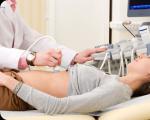
The procedure itself cannot be called uncomfortable: the patient sits on the tomograph couch, and a scanner circles around him, taking pictures. All that is required of the subject is to remove objects made with metal inclusions (hairpins, piercings, brassiere with metal underwires, etc.) and lie still. The duration of the procedure is about 15 minutes, and with the introduction of contrast it takes about half an hour. The conclusion in most cases is ready 2-3 hours after the end of the study.
What can be revealed on CT images of the abdominal cavity and retroperitoneum?
CT results help the doctor evaluate the functionality of the internal organs and tissues of the abdominal cavity and retroperitoneal space, as well as the effectiveness of the chosen treatment. CT can also reveal:

Features of SCT of the abdominal cavity with bolus contrast
The drug can be administered intravenously, orally or rectally. To examine the upper gastrointestinal tract, the patient is asked to drink a special liquid. In order to contrast the large intestine, an enema with a contrast agent is used. The bolus method is used to visualize the abdominal organs.
Computed tomography with bolus contrast is characterized by the introduction of a special substance by an automated injector at a programmed speed and time of delivery of the drug. In this case, the doctor conducting the study must take into account the clinical task, the number of years and body weight of the person, as well as other features.
Bolus contrast allows one to clearly identify and delimit tumor neoplasms, assess the extent of spread of a malignant tumor and its resectability, and identify metastases in lymph nodes and parenchymal organs.
Information content of CT without contrast

Unlike CT without contrast, a study with the introduction of a special drug allows you to most accurately determine the condition of soft tissues, examine the arterial and venous beds, examine the vessels supplying blood to the kidney tissue, gastrointestinal tract organs and other internal organs. The use of a contrast agent allows you to study the lymphatic system, examine the rectum and colon, and also carefully examine any parenchymal area.
Consequences and complications after computed tomography
An iodine-based drug is used as a contrast agent. After entering the blood, the accumulation of contrast stains the tissue, and thereby displays the area being studied in the images. Contrast drugs are completely eliminated from the patient’s body within 48 hours, and their volume depends on the body weight of the patient. To quickly remove the contrast, doctors recommend drinking as much fluid as possible.
The examination of the abdominal cavity itself does not cause serious complications or side effects. However, a number of patients complain of dizziness or attacks of nausea when rotating the scanner, similar to the state of motion sickness on a carousel in a theme park. After the procedure is completed, the unpleasant sensations disappear.
When using the bolus diagnostic method, people with sensitive skin may experience itching or notice redness at the site of the needle prick. When administering the drug orally, a taste of iodine may appear.
Also, do not worry if the patient feels cold or hot during the contrast injection. These signs do not require the intervention of a medical professional and will go away on their own.
A more negative effect of CT on the patient’s body may appear when the person did not know that he was suffering from an allergy to iodine. In this case, the help of a doctor and the use of antihistamines are necessary. If the patient begins to experience difficulty breathing, a cough appears, or a rash and swelling develop on the skin, you must immediately inform the doctor.
Cost of examination
The price for examining the abdominal cavity and retroperitoneal space depends on the area being examined. The more images required for diagnosis, the higher the cost. Computed tomography without contrast is cheaper, since a special substance is not required to be injected into the patient's body. On average, the price of chest diagnostics using CT in Moscow clinics starts from 4 thousand rubles.
A CT scan of the abdominal organs will provide the specialist with comprehensive information about the condition of the internal organs - liver, pancreas, lymph nodes, etc. The article describes how the examination is carried out, what needs to be done before a CT scan of the abdominal cavity as preparation, what are the indications and contraindications for the procedure.
CT scan of the entire abdominal cavity is a highly informative diagnostic procedure that can show an accurate three-dimensional image of parenchymal and hollow organs, vessels and the lymphatic system of a given anatomical zone. This method involves the use of small doses of X-ray irradiation, giving minimal radiation exposure, while allowing to obtain layer-by-layer sections of tissue with a pitch of 0.5-10 mm, depending on the type of tomograph.
CT is indispensable in cases of suspected oncology, acute and chronic diseases of the peritoneal organs and blood vessels, because the accuracy of the study is much higher than that of ultrasound and radiography. What does this procedure show, what organs are visible during it? This:
- Organs related to the gastrointestinal tract system - large, small intestine (overview), pancreas.
- The organs of the hepatobiliary system are the liver, gallbladder and ducts.
- Spleen.
- The organs of the retroperitoneal space are lymph nodes and ducts, kidneys and adrenal glands.
- Vessels of the abdominal cavity and retroperitoneal space.
Nowadays, CT of the abdominal organs is often replaced by MSCT, an even more modern research method. It is a multislice computed tomography, which is favorably distinguished by its technical capabilities and the number of sections when scanning organs. The image quality during this examination is higher, the execution time is shorter, and the radiation dose is lower.
Why do you need a CT scan with contrast?

The usual tomography procedure (native CT) is performed without the introduction of a contrast agent. But often more accurate data is needed, and this is possible using contrast-enhanced abdominal CT. It is performed during intravenous urography, or the introduction of a special substance into the body that colors the organs in a contrasting shade.
During urography, the area under study will acquire an uneven color: pathologically altered tissues will differ sharply from healthy ones in color in the pictures. Thus, urography during abdominal CT with contrast increases the accuracy and reliability of the data obtained.
Typically, an iodine-based drug (Omnipak, Urografin) is used as a contrast for CT urography, so the possibility of allergic reactions to these substances should be taken into account. Urografin and other drugs give side effects (nausea, hyperthermia, chills, rash, anxiety, etc.) only in 1% of cases. Before using Urografin, it is recommended to conduct a special test - administering a small dosage of the drug subcutaneously before performing tomography.

Contrast agent for peritoneal tomography can be administered in different ways:
- Intravenous - used as a single injection, it allows you to clearly visualize the condition of blood vessels and hollow organs.
- Oral – helps check the health of the liver, pancreas, kidneys, that is, parenchymal organs (they consist of solid tissue).
- Rectal - more often used to assess the condition of the large intestine.
- Bolus – used when a detailed examination of many organs is necessary; it involves drip intravenous administration of a contrast agent using a special device – an automatic injector.
The dose of Urografin is calculated individually based on the patient’s weight. Pictures are usually taken 3-12 minutes after the drug is administered (depending on the organ being examined).
Indications for examination of the peritoneum

It is recommended to perform this procedure if any neoplasm is suspected - benign (polyps, cysts, fibromas, lipomas, adenomas, etc.), malignant (tumors of the intestines and internal organs). A CT scan will show the size and boundaries of tumors, the presence of metastases, and the degree of growth into the walls of organs.
The list of indications for CT is very extensive. Tomography is indispensable for inflammatory processes in the abdominal cavity, dropsy (ascites), traumatic injury, for example, closed (blunt) abdominal trauma. The method is excellent for diagnosing the condition of the liver and identifying cirrhosis and its stage, acute and chronic hepatitis. Reflects CT and diseases of the pancreas, in particular, diabetic changes, pancreatitis.

Emergency conditions accompanied by an acute abdomen (appendicitis, abdominal abscesses, hernias with torsion and necrosis, etc.) will also be visible on computed tomography images. The examination is also indicated for:
CT is also performed if magnetic resonance imaging is contraindicated for a patient for some reason, but there is a need for such an accurate diagnosis. Also, a CT scan will show all anomalies and deviations from the norm in the location of congenital organs, which may be necessary for both a child and an adult. Symptoms for which a doctor may recommend diagnostics:
- Yellowing of the skin
- Stomach ache
- Weight loss
- Chronic digestive disorders
- Problems with urine output
- Signs of hemorrhage
How to prepare for an abdominal CT scan?

Measures to prepare for this procedure are extremely important to obtain an accurate result. Preparation for a CT scan of the abdominal cavity is carefully planned, and all doctor’s recommendations are followed. The procedure is performed strictly on an empty stomach, since the digestive organs can change shape and volume after eating or drinking a large amount of water. It is also important to prevent increased gas formation, so as not to distort the picture of the images.
To make intestinal motility less intense, and also to free it from stagnant feces, you should start following a special diet 2-3 days before the examination. Removed from the menu:
- Peas, beans, corn, apples, rye bread, cabbage of any kind, baked goods, nuts, prunes are products that increase gas formation.
- Pastries and cream cakes, strong coffee, tea, milk, pasta and other foods that cause constipation.
Also, you should not eat a lot of vegetables and fruits that have not been cooked. It is advisable to eat lean meat, steamed or boiled, stewed vegetables, unleavened cottage cheese, dried white bread, boiled or stewed fish.

The CT scan itself is performed on an empty stomach, and at least 8 hours must have passed since the last meal. When the diagnosis is scheduled for the morning, only a light dinner is taken in the evening. If the procedure is planned in the afternoon or evening, then in the morning you can take some light food.
As prescribed by the doctor, a cleansing enema is given before the procedure, or a solution of the drug Fortrans is taken in the evening to cleanse the intestines. This is especially necessary if a thorough examination of the colon is required. During menstruation or pathological gas formation, enterosorbents or carminatives are taken before the study.
When preparing for an abdominal CT scan with contrast, you should consider all the same recommendations that have already been listed. You need to tell the specialist about all the medications you are taking, the presence and types of allergies, and chronic diseases. Sometimes, before a CT scan with contrast, it is recommended to take a biochemical blood test and show it to a radiologist.
Directly in the office you need:
- Remove all metal jewelry and watches.
- Empty pockets.
- Remove removable dentures.
- Wear loose clothing.
How is the procedure performed?

Before starting the examination, it is important to transfer to the specialist all previous transcripts of instrumental studies, as well as protocols of previously performed CT scans of the abdominal cavity. Next, the patient is injected with a contrast agent or allowed to take it by mouth (if required). After a certain period of time, the person is placed on a couch or a special pull-out table.
The patient is pushed onto the table into the tomograph tunnel. The doctor leaves the room with the tomograph, but can communicate with the person being examined through a microphone. The device takes a series of pictures. It is important to lie still during the diagnosis to ensure accurate results. Sometimes patients are asked to hold their breath for a short period of time.
The whole procedure takes 30-60 minutes.
The price of a computed tomography scan of the abdominal organs will depend on the complexity, location of the pathological focus, the presence or absence of contrast, the type of administration and the amount of contrast agent. The price can vary from 3500 to 7000 rubles.
Contraindications to abdominal tomography

Although safe, CT scans involve the use of X-rays and are therefore contraindicated during pregnancy. Relative contraindications are:
- Age up to 7 years
- Acute phase of the inflammatory process in the kidneys
- Lactation (you cannot breastfeed for 24 hours after diagnosis)
- Large patient weight (limitation depends on the capabilities of the device)
- Hyperkinesis, mental illness (examination with sedation is possible)
Contrast with Urografin or other iodine-based agents is contraindicated in the following cases:
- Severe diabetes mellitus
- Allergies to contrast media
- Serious diseases of the liver, kidneys
- Taking incompatible medications
- Hyperthyroidism
- Decompensated heart failure

Diseases for which computed tomography of the abdominal cavity is prescribed:
- Lymphadenitis
- Abscess
- Vascular atherosclerosis
- Aneurysm
- Kink of blood vessels
- Thrombosis
- Cirrhosis
- Hepatitis
- Fatty liver hepatosis
- Urolithiasis disease
- Echinococcosis
- Cholelithiasis
- Hemangioma
- Carcinoma and other types of cancer of the kidneys, pancreas and other organs
- Lymphoma
- Hemochromatosis
- Hydronephrosis
- Pheochromocyotoma
- Polycystic kidney disease
- Nephroptosis
- Appendicitis
- Diverticulosis
- Cholangitis
- Pancreatitis
CT scan results are accurate and allow early diagnosis. But without specific indications, it is better not to overuse CT, because a number of diseases can be diagnosed using cheaper and simpler methods.
Abdominal CT (computed tomography of the abdominal cavity) is a technique that creates images of internal organs using X-rays in the form of slices in order to detect early signs of disease. Diagnostics using CT allows you to assess the condition of the abdominal cavity, determine the stages of the disease and build a further treatment plan. In certain cases, for better visualization of the studied structures, a CT scan of the abdominal cavity with contrast is performed. A CT scan of the abdominal cavity can be performed on a child only if there are absolute indications and other diagnostic methods are not informative. A tomogram of the abdominal cavity in children from the moment of birth should be performed only if the benefit from the study is higher than the harm from radiation.
contrast study,
Indications
Indications for CT scan of the abdominal cavity: closed abdominal trauma, detection of damage to internal organs; cystic formations and tumors of the abdominal organs; metastases to deep lymph nodes; abscesses of the abdominal organs and the spaces between them; vascular disorders leading to secondary changes in organs; diffuse liver diseases; obstructive jaundice; hepatosplenomegaly; contraindications to MRI.
Preparation
Patients with diabetes and taking glucophage must obtain permission from the referring physician before testing. During a contrast study, an intravenous injection of contrast material is given, so refrain from eating or drinking water for 4 hours before the test. The main contraindications for abdominal CT include: pregnancy and lactation, weight more than 150 kg, intolerance to contrast, unstable mental state of the patient, the presence of metal objects (implants) in the body.
More details
Price
The cost of an abdominal CT scan in Moscow ranges from 2,500 to 25,900 rubles. The average price is 7240 rubles.
Where can I get an abdominal CT scan?
Our portal contains all the clinics where you can get an abdominal CT scan in Moscow. Choose a clinic that suits your price and location and make an appointment on our website or by phone.
The study of the abdominal organs, until today, has had rather limited possibilities. The use of radiography made it possible to obtain only overview images, with the help of which it was possible to evaluate the stomach and duodenum, isolated using barium contrast. An image of the gallbladder was obtained using cholecystography. The greatest difficulty was the diagnosis of diseases of parenchymal organs, the structure of which was practically not determined on a regular x-ray.
The emergence and development of computerized systems (tomographs), which make it possible to process the information obtained as a result of scanning in a short period of time, has significantly improved the quality of diagnostics. Computed tomography of the abdominal cavity has opened up great opportunities for direct and indirect assessment of the condition of the liver, gallbladder and its ducts, spleen and pancreas.
How CT works
Computed tomography is a study, the essence of which is to scan the patient’s body with a fan beam of X-ray radiation, while simultaneously recording the degree of its attenuation after overcoming obstacles in the form of organs and tissues. At the same time, the tube and recording detectors rotate 360°, recording the received data every 1–3°.
Depending on the generation of the tomograph used for diagnosing abdominal organs (AOC), it may have the following design features:
- registration of residual radiation is carried out by one row of detectors rotating simultaneously with the X-ray tube;
- rotation is carried out only by the X-ray tube, and the attenuation of radiation is recorded by stationary sensors located along the entire perimeter of the circle;
- when the emitter is continuously rotated, there is a simultaneous translational movement of the table with the patient lying on it (spiral CT);
- The mechanism of spiral CT (rotation of the emitter and step-by-step movement of the table) is supplemented by an increase in the number of rows with recording detectors (MSCT). Multislice computed tomography allows you to increase the number of optical sections obtained simultaneously.
Important! The latest achievement in the development of CT is a tomograph with 320 rows of detectors, which allows one to simultaneously obtain 320 sections, that is, a layer-by-layer image of one organ, per revolution of the emitter.
Main tasks of CT
In addition to X-ray computed tomography, ultrasound, magnetic resonance imaging (MRI) and positron emission tomography (PET) are used to diagnose ABP pathologies. At the same time, the last two methods differ only in the way of obtaining the initial information; computer processing of the received data is carried out according to the same principles. The diagrams below show that the transducer, computer, and display are an integral part of X-ray CT and MRI.
Structural diagram of a magnetic resonance imaging scanner, which differs from the operation of CT radiation
The main task of tomography when examining OBP is to determine their following parameters:
- form;
- location;
- dimensions;
- clarity of contours;
- the presence of pathological foci;
- intensity of the shadow of pathological foci;
- difference in tissue density of the organ under study and the pathological formation.
Determination of density using tomography is carried out by mathematical processing of data on the degree of absorption of X-ray radiation (for X-ray CT) and the intensity of electromagnetic radiation (for MRI). The result of a computer reconstruction of the obtained data is displayed on the monitor in the form of an image where the organs, depending on their density, have the appearance of shadows of varying degrees of severity.
A characteristic phenomenon when studying ABP using CT is their insufficient visualization, which is associated primarily with the specific structure of the parenchyma of the liver, pancreas and spleen. However, if on a regular X-ray, the shadow may not be sufficiently pronounced, and it is not possible to get a complete picture of the state of the organ being examined, then with CT, even minor changes in the signal allow you to see a fairly clear image.
Indications
Despite the great potential of CT in diagnosing diseases of the chest organs, the technique is not a priority in the study of ABP. This is partly due to the limited capabilities of a computer monitor, which is unable to reflect all shades of gray, which as a result significantly reduces the information content of the examination.
In this regard, computed tomography of the abdominal organs is mainly used in the differential diagnosis of neoplasms. The value of tomography in detecting malignant and benign neoplasms of the pancreas and liver, as well as in identifying metastases, cannot be underestimated.
In this regard, the main indications for CT scan of the liver, pancreas and spleen are:
- suspicions of the presence of primary or secondary malignant neoplasms;
- detection of space-occupying formations (hydatid cyst, adenoma);
- abscess;
- cirrhosis of the liver;
- suspicion of the presence of stones in the gallbladder or ducts;
- acute pancreatitis;
- ischemic tissue damage to parenchymal organs, leading to necrosis;
- a history of cancer;
- to differentiate the diagnosis of abdominal pain of unknown origin.

Carrying out an ultrasound scan before a CT scan will help to obtain the most informative diagnostic picture.
CT with contrast
It is possible to achieve the maximum level of information content when performing CT scans of the abdominal cavity and retroperitoneal space using contrast enhancement. The use of X-ray contrast agents is especially important for the early detection of cancer of the liver and pancreas and the diagnosis of vascular pathologies leading to tissue damage.
When performing a conventional (native) CT examination, you can only get a general idea of the state of the organ, for example, identify anatomical deviations (increase or decrease in size, incorrect location), detect foci of calcification, indicating oncological or inflammatory processes in the tissues of the organ, determine the presence of stones in the gallbladder or bile ducts.
Improving the quality of the information obtained through the use of contrast is based on improving the visualization of the circulatory system in the vascular system, and directly in the tissues of organs.
After introducing contrast into a vein, you can get 2 types of changes in the tomographic picture: vascular, parenchymal. The first change occurs immediately after administration of the drug, and the second as the contrast agent accumulates and distributes in the tissues.
By the rate and intensity of accumulation of the drug by the parenchyma, one can judge the presence of a neoplasm and its differential affiliation (malignant, benign, cystic). The presence in the liver or pancreas of a strongly pronounced vascular network or a focus with an increased concentration of contrast agent indicates the malignant nature of the neoplasm. A uniform distribution of contrast in tissues, even when an enlarged node or organ deformation is detected, may indicate benign tissue growth (adenoma).
Cysts, necrotic areas and cavities are defined as areas free of contrast. To highlight hollow organs (stomach, duodenum, intestines) with an X-ray contrast agent, as well as to improve visualization of the pancreas, barium sulfate is used. The water-soluble suspension is drunk and, distributed in the body, it envelops the walls, forming clear contours of the gastrointestinal tract in the image, allowing one to evaluate their shape and the integrity of the walls.
Important! Due to the peculiarities of the structure and location of the pancreas, which has slight differences in tissue density, its best visualization can be achieved using a barium contrast stain of the stomach.
Preparation
Preparation for a CT scan of the abdominal cavity and pelvis includes refusing food that can stimulate gas formation in the intestines 2–3 days before the procedure and completely refusing food 7–9 hours before the diagnosis. Diagnosis of the gastrointestinal tract with contrast enhancement requires a longer fast (about 12 hours) and preliminary cleansing of the intestines using laxatives and enemas.
Due to the fact that to perform a CT scan it is necessary to remain completely still for a certain period of time, anesthesia is used to examine newborns and young children whose actions and movements cannot be controlled. If, in the presence of one of the parents, the child is able to lie still for 20–25 minutes, his presence in the immediate vicinity of the tomograph is allowed, provided that X-ray protective equipment is used (lead apron). Bowel preparation is a prerequisite for diagnosing ABP, since the formation of gases can lead to distortion of the image, and, accordingly, to difficulties in deciphering the results.

Drinking carbonated water can cause gas in the intestines
Carrying out
A CT scan of the PD organs is carried out in the same way as a tomography of any other area, with the only difference being that the scan is carried out in the abdomen. The patient lies down on the couch, raises his arms up and the table takes the position corresponding to the beginning of the study area. To obtain a layer-by-layer image of all OBPs, scanning begins from the upper dome of the diaphragm.
The duration of the procedure depends on the type of equipment used (number of sections obtained simultaneously), table pitch and the need to use contrast. The examination tactics are determined by the doctor immediately before diagnosis, based on the medical history and the available results of a previous examination (laboratory tests, ultrasound data).
CT with contrast takes longer, especially when examined on older equipment, where the drug is injected manually and scanning is done over a long period to cover the entire time period that the process of contrast distribution in tissues takes. Modern tomographs automatically introduce a contrast agent, and in accordance with the expected phases of distribution of the drug in the tissues, scanning is carried out.
This tactic makes it possible to take into account not only the degree of increase in the concentration of the drug in tissues, but also the rate of its distribution. It is known that malignant neoplasms accumulate radiopaque substances to a greater extent than healthy tissues, but the accumulation process occurs much more slowly.
Tomographic picture
To get a complete picture of what CT shows and how great its diagnostic capabilities are, it is necessary to consider the most common pathologies of the liver and pancreas, from the point of view of the information content of tomography.
On conventional tomographic images, a healthy liver has smooth, well-defined edges, uniform density (+60 to +70 Hounsfield scale) and uniform structure. Against the background of the parenchyma, liver vessels are clearly visible, having a lower density (from +30 to +50 on the Hounsfield scale). The internal structure is assessed depending on where the cut is made.
The main pathological changes in the liver include:
- fatty hepatosis. When diagnosing fatty hepatosis, CT is considered the most informative method, since with an increase in the amount of adipose tissue, the density of the liver decreases significantly. The tomogram clearly shows areas of reduced density (fatty infiltrates) surrounded by healthy tissue. With complete damage to the liver, its increase and decrease in density are observed. If a healthy organ has a density 8 Hounsfield units (HU) more than the spleen, then the fatty areas have a significantly lower density;
- chronic hepatitis. It is impossible to make a diagnosis of hepatitis on the basis of a CT scan alone, since during the scan only diffuse enlargement of the organ is determined, the density of the parenchyma remains normal. However, when using a positron emission tomograph (PET) using radioisotope drugs, a decrease in the functional activity of the liver becomes obvious, which is a sign of sclerosis of liver cells;
- cirrhosis A disease accompanied by wrinkling and scarring of liver tissue. The initial stages of the disease are characterized by an increase in the size of the liver and spleen, which is caused by increased pressure in the veins; varicose veins are often observed. In the later stages, the organ shrinks, it decreases in size, blurred contours appear, and nodes form. The parenchyma on the tomogram looks heterogeneous, with dense fatty inclusions.

The tomogram shows heterogeneity of the parenchyma, characteristic of liver cirrhosis
When examining the pancreas, the most common pathology is pancreatitis, manifested on an X-ray tomogram by the following signs:
- loss of clarity of contours;
- decrease in parenchyma density of the entire organ or focal;
- swelling of surrounding tissues;
- reduced accumulation of contrast agent;
- the appearance of necrotic zones that do not accumulate the contrast agent.
When a tumor is detected, deformation of the contour of the gland and increased accumulation of contrast agent by the neoplasm are observed. This allows you to get an idea of the structure, size and contours of the tumor. Detection of malignant lesions of lymph nodes in the retroperitoneal space is based on an assessment of their size and number. Even with normal size of lymph nodes, an increase in their number usually indicates the spread of malignant metastases.
Despite the undoubted effectiveness of CT in diagnosing AKD diseases, this method cannot be considered universal. The disease of each organ requires the selection of diagnostic techniques, the order of which depends on the severity of the process and clinical manifestations. If ultrasound can give a general definition of the nature of the lesions, then to make an accurate diagnosis, it is advisable to use CT.